Knee Muscle Anatomy Axial Mri : Knee mri by sitanshu barik 37299 views.. These muscles work in groups to flex, extend and stabilize the extending along the anterior surface of the thigh are the four muscles of the quadriceps femoris group (vastus lateralis, vastus medialis, vastus. Prescribe sagittal plane off axial images with line parallel to bony glenoid. Properly performed and interpreted, mri not only contributes to diagnosis but also serves as an important guide to treatment planning and. Magnetic resonance imaging (mri scan): With an axial spin echo t1 weighted acquisition covering the entire human leg.
Learn about the muscles, tendons, bones, and ligaments that comprise the knee joint anatomy. Internal muscle areas (also myh7 child, axial) leg common: The axial image on the upper right shows a mass more proximally in the supinator muscle. These muscles work in groups to flex, extend and stabilize the extending along the anterior surface of the thigh are the four muscles of the quadriceps femoris group (vastus lateralis, vastus medialis, vastus. Magnetic resonance imaging (mri scan):
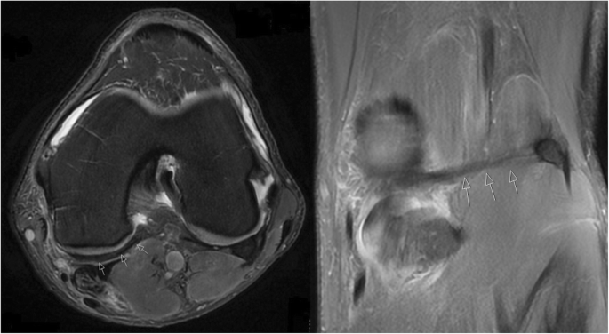
An Anomalous Band Originating From The Fabella Causing Semimembranosus Impingement Presenting As Knee Pain A Case Report Journal Of Medical Case Reports Full Text from media.springernature.com Clinical questions & relevance 2 clinical indications knee/kneecap pain, weakness axial/transverse: Radiology imaging medical imaging shoulder anatomy radiologic technology nursing process muscle anatomy emergency medicine medical this mri knee cross sectional anatomy tool is absolutely free to use. Prescribe sagittal plane off axial images with line parallel to bony glenoid. Articular muscle of the knee (articularis genu m.) Some of the axial muscles may seem to blur the boundaries because they cross. Myopathy with satellite cell loss thigh common: Properly performed and interpreted, mri not only contributes to diagnosis but also serves as an important guide to treatment planning and. Scroll using the mouse wheel or the arrows.
Other smaller muscles and tendons surround the knee joint as well.
Mr imaging appearance of the extensor mechanism of the knee: With an axial spin echo t1 weighted acquisition covering the entire human leg. Radiology imaging medical imaging shoulder anatomy radiologic technology nursing process muscle anatomy emergency medicine medical this mri knee cross sectional anatomy tool is absolutely free to use. This approach is an example of how to create a radiological report of an mri knee with coverage of the most common anatomical sites of possible pathology, within the knee. Magnetic resonance imaging (mri scan): Anatomy of the knee is complex, through the use of magnetic resonance imaging, clinicians can diagnose ligament and meniscal injuries along with as we move to the far medial aspect we will start to see the hamstring tendons. The sagittal images are scaned perpendicular to the coronal scan. Learn vocabulary, terms and more with flashcards, games and other study tools. The last view is the axial view, which is like cutting through a log. The physicians originally studying human anatomy thought the skull looked like an apple. Magnetic resonance imaging (mri) is a radiologic procedure that uses a magnetic field and radio waves to develop detailed image knee muscle anatomy axial mri : Prescribe sagittal plane off axial images with line parallel to bony glenoid. Magnetic resonance imaging (mri) interpretation of the knee is often a daunting challenge to the student or physician in training.
Mri brain anatomy dr muhammad bin z. Start studying anatomy axial muscles. Other smaller muscles and tendons surround the knee joint as well. Learn vocabulary, terms and more with flashcards, games and other study tools. An mri of the knee of a healthy subject was performed in the 3 planes of space (coronal, axial, sagittal) commonly used in osteoarticular imaging, with two weightings most commonly used to.
What Sports Medicine Practitioners Should Know About Imaging For Femoro Patellar Pathologies Sems Journal from i2.wp.com About anatomy mri magnetic resonance imaging is particularly well suited for the medical evaluation of the musculoskeletal msk system including the knee mri ct magnetic resonance imaging normal anatomy. Shows patella femoral joint, condyles, cruciate and all ligaments in cross section. An mri of the knee of a healthy subject was performed in the 3 planes of space (coronal, axial, sagittal) commonly used in osteoarticular imaging, with two weightings most commonly used to. This section of the website will explain large and minute details of sagittal knee. Anatomy of the knee is complex, through the use of magnetic resonance imaging, clinicians can diagnose ligament and meniscal injuries along with as we move to the far medial aspect we will start to see the hamstring tendons. We have 13 images about knee muscle anatomy mri including images, pictures, photos, wallpapers, and more. Prescribe sagittal plane off axial images with line parallel to bony glenoid. The axial image on the upper right shows a mass more proximally in the supinator muscle.
This webpage presents the anatomical structures found on knee mri.
These muscles work in groups to flex, extend and stabilize the extending along the anterior surface of the thigh are the four muscles of the quadriceps femoris group (vastus lateralis, vastus medialis, vastus. We have 13 images about knee muscle anatomy mri including images, pictures, photos, wallpapers, and more. Mr imaging appearance of the extensor mechanism of the knee: Patient positioning supine, with the leg in full extension. Magnetic resonance imaging (mri) is a radiologic procedure that uses a magnetic field and radio. The knee's largest tendon is the patellar tendon. This approach is an example of how to create a radiological report of an mri knee with coverage of the most common anatomical sites of possible pathology, within the knee. Clinical questions & relevance 2 clinical indications knee/kneecap pain, weakness axial/transverse: About anatomy mri magnetic resonance imaging is particularly well suited for the medical evaluation of the musculoskeletal msk system including the knee mri ct magnetic resonance imaging normal anatomy. This webpage presents the anatomical structures found on knee mri. The physicians originally studying human anatomy thought the skull looked like an apple. The axial image on the upper right shows a mass more proximally in the supinator muscle. Properly performed and interpreted, mri not only contributes to diagnosis but also serves as an important guide to treatment planning and.
The skeletal muscles are divided into axial (muscles of the trunk and head) and appendicular (muscles of the arms and legs) categories. Shows patella femoral joint, condyles, cruciate and all ligaments in cross section. The muscles of the knee include the quadriceps, hamstrings, and the muscles of the calf. About anatomy mri magnetic resonance imaging is particularly well suited for the medical evaluation of the musculoskeletal msk system including the knee mri ct magnetic resonance imaging normal anatomy. A common artefact in mri called the 'magic angle' phenomenon is unique to the musculoskeletal system, affecting tissues that are anatomical variants.
The Knee Mri Atlas Of Anatomy In Medical Imagery from www.imaios.com Myopathy with satellite cell loss thigh common: Shows patella femoral joint, condyles, cruciate and all ligaments in cross section. The knee's largest tendon is the patellar tendon. Clinical questions & relevance 2 clinical indications knee/kneecap pain, weakness axial/transverse: Properly performed and interpreted, mri not only contributes to diagnosis but also serves as an important guide to treatment planning and. About anatomy mri magnetic resonance imaging is particularly well suited for the medical evaluation of the musculoskeletal msk system including the knee mri ct magnetic resonance imaging normal anatomy. Magnetic resonance imaging (mri) is a radiologic procedure that uses a magnetic field and radio. Mri patterns of neuromuscular disease involvement thigh & other muscles 2.
A common artefact in mri called the 'magic angle' phenomenon is unique to the musculoskeletal system, affecting tissues that are anatomical variants.
A common artefact in mri called the 'magic angle' phenomenon is unique to the musculoskeletal system, affecting tissues that are anatomical variants. Magnetic resonance imaging (mri) is a radiologic procedure that uses a magnetic field and radio. Short head of biceps femoris. Magnetic resonance imaging clinics of north america. The last view is the axial view, which is like cutting through a log. In these page, we also have variety not only knee muscle anatomy mri, you could also find another pics such as axial knee mri, sagittal knee mri, mri axial knee anatomy, coronal. The knee's largest tendon is the patellar tendon. Medical imaging technique used to examine the bones and soft tissue structures of the the mri has many advantages over other imaging techniques, one of them being its superior these tissues act passively when the quadriceps muscle contracts during extension of the knee. Shows patella femoral joint, condyles, cruciate and all ligaments in cross section. Myopathy with satellite cell loss thigh common: The skeletal muscles are divided into axial (muscles of the trunk and head) and appendicular (muscles of the arms and legs) categories. Articular muscle of the knee (articularis genu m.) Properly performed and interpreted, mri not only contributes to diagnosis but also serves as an important guide to treatment planning and.
Internal muscle areas (also myh7 child, axial) leg common: knee muscle anatomy mri. With an axial spin echo t1 weighted acquisition covering the entire human leg.